In the development of new materials and in industrial quality control, accurately understanding what a surface looks like at the microscopic scale can make the difference between a product that meets specifications and one that fails in service. Roughness, porosity, surface defects, or the condition of a coating are parameters that often cannot be assessed with the naked eye or with conventional measurement instruments.
This is where advanced optical characterization techniques come into play, particularly confocal microscopy, one of the most versatile tools for obtaining high-resolution images and topographic data without physical contact or damage to the sample. In this article, we explain what confocal microscopy is, how it works, the different types available, its main applications, and how it relates to material surface topography and the equipment used to characterize it.
What is confocal microscopy?
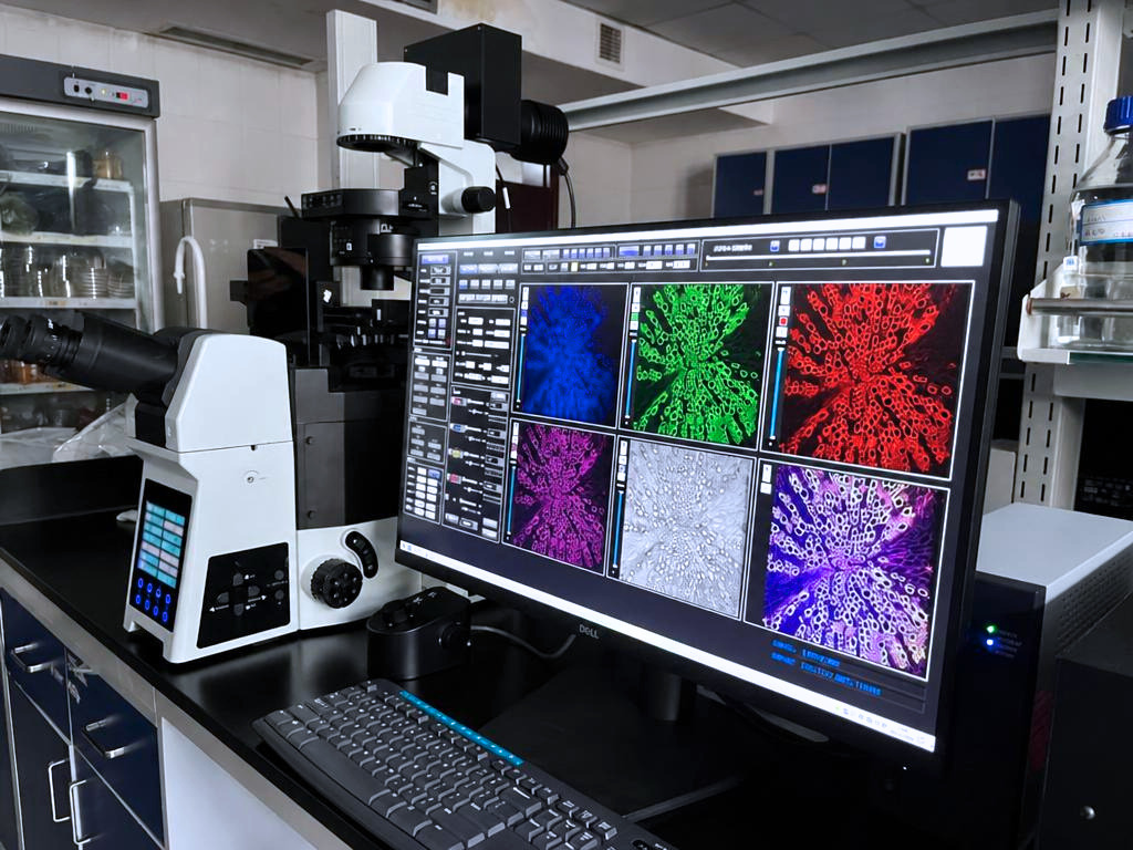
Confocal microscopy is an optical microscopy technique that enables high-resolution, highly detailed images to be obtained at different depths within a sample by eliminating out-of-focus light originating from planes outside the focal region.
Unlike a conventional optical microscope, which simultaneously captures light from a broader volume of the sample, a confocal system illuminates and observes a specific point at a time. This makes it possible to improve image contrast and definition, both laterally and in depth.
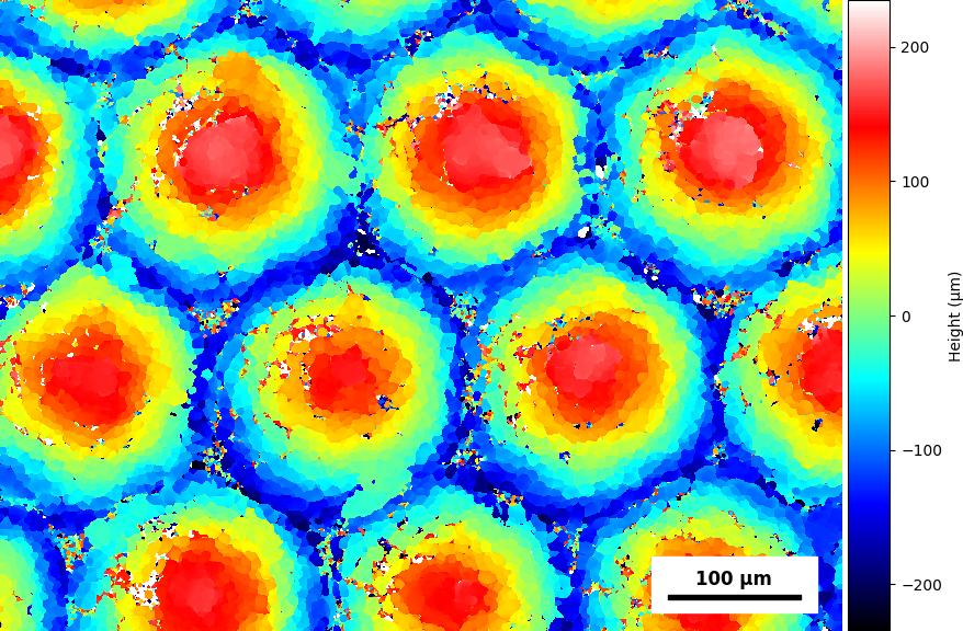
The ability to obtain information from different focal planes makes it possible to perform an “optical sectioning” of the sample and, by combining the different depth levels, reconstruct the surface in three dimensions. For this reason, it is particularly useful for studying the microstructure and topography of materials with a high degree of precision.
How does a confocal microscope work?
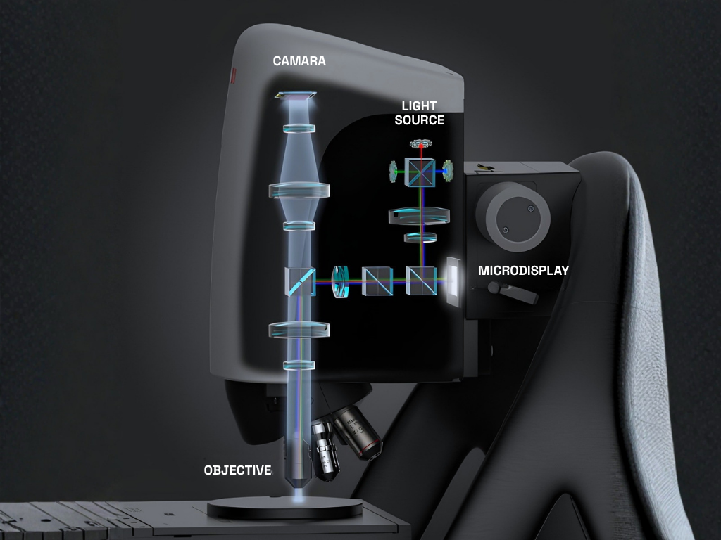
The operating principle of a confocal microscope is based on a point illumination system, generally using a laser, and on a diaphragm or aperture known as a pinhole, positioned in a plane conjugate to the focal plane of the sample.
The pinhole is a key component of the system, as it allows mainly the light originating from the focused plane to reach the detector, while blocking most of the scattered light coming from other planes.
In simplified terms, the process follows these steps:
- A beam of light, usually a laser, is focused onto a specific point on the sample.
- The light reflected or emitted by that point passes through the optical system and through the pinhole, which filters out signals from out-of-focus areas.
- A detector records the intensity of the light coming from the point being analyzed.
- The system scans the surface, successively recording different points to build the image.
- The software processes the acquired information and generates a digital image. By combining data corresponding to different depths, it is possible to obtain a three-dimensional reconstruction of the surface.
This procedure makes it possible to obtain not only highly detailed images, but also information about the height and relief of the surface, which can be used to generate profiles and topographic maps.
Types of confocal microscopy
There are different configurations of confocal microscopy, adapted to the characteristics of the sample and the objectives of the analysis. The most common ones are:
- Confocal Laser Scanning Microscopy (CLSM): Uses a laser to scan the sample point by point. Thanks to a pinhole system, most of the light originating from out-of-focus planes is eliminated, resulting in images with higher contrast and resolution than conventional microscopy. In addition, it allows optical sectioning at different depths and three-dimensional reconstruction of samples. It is widely used in cell biology, histology, and the analysis of microscopic structures.
- Spinning Disk Confocal Microscopy: Uses a disk containing multiple microlenses and pinholes, allowing numerous points of the sample to be illuminated and detected simultaneously. This considerably increases the acquisition speed and reduces the sample’s exposure to light, thereby minimizing the risk of phototoxicity and photobleaching. Therefore, it is particularly suitable for observing dynamic processes in living cells, such as organelle movement, cell division, or intracellular transport.
- Wide-Field Confocal Microscopy: Seeks to achieve a balance between acquisition speed and image quality, allowing relatively large areas of the sample to be analyzed in a short period of time. Unlike point-scanning systems, it can acquire a greater amount of information simultaneously. This makes it useful when studying large samples or performing rapid observations, although the level of resolution and out-of-focus light rejection may vary depending on the system used.
- Fluorescence Confocal Microscopy: Analyzes the light emitted by fluorophores present in the sample after they are excited at a specific wavelength. It allows specific molecules or structures to be identified and localized through the use of fluorescent markers, such as proteins, antibodies, or probes. It is particularly useful in biological and biomedical applications, as it enables the study of the distribution and localization of different cellular components and, through the use of different fluorophores, the simultaneous analysis of several structures.
Applications of confocal microscopy
The versatility of this technique allows it to be used in numerous fields, ranging from biological research to industrial surface characterization. Its main applications include:
- Health sciences and biology: Study of tissues, cells, and subcellular structures.
- Materials engineering: Characterization of the microstructure of metals, polymers, ceramics, and composite materials, as well as the analysis of coatings and thin films.
- Industrial quality control: Inspection of machined surfaces, detection of defects, cracks, or porosity, and verification of the surface finish of manufactured components.
- Roughness and topography analysis: Measurement of height profiles and surface roughness.
- Semiconductors and microelectronics: Inspection of wafers, circuits, and micrometer-scale components where high resolution is required.
In industrial applications, these capabilities make confocal microscopy particularly valuable when it is necessary to combine visual information with quantitative data about the surface.
What is material surface topography?
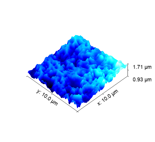
Material surface topography involves studying and characterizing the irregularities, relief, and finish of a surface at different scales, from the macroscopic to the micro- and nanometric levels. Parameters that can be analyzed include roughness, waviness, peak and valley heights, surface porosity, and the presence of defects, cracks, or scratches.
Its characterization is essential in numerous industrial processes, as the condition of a surface can directly influence properties such as friction, wear, coating adhesion, corrosion resistance, optical behavior, and the final aesthetic appearance.
For this reason, topographic analysis is part of different stages of product development, manufacturing process validation, and quality control.
Material topography equipment and testing
There are different types of equipment and techniques for measuring and characterizing the topography of a material. The choice depends on factors such as the required resolution, the size and characteristics of the surface, and whether or not contact with the sample is required.
The most widely used systems include:
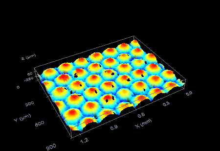
- Confocal microscopes: enable detailed topographic maps to be obtained without contact, combining image and height information in a single analysis.
- Optical profilometers and interferometers: determine height variations by analyzing light and interference patterns, providing accurate measurements of parameters such as roughness and flatness.
- Contact profilometers (roughness testers): use a stylus or probe that physically traverses the surface to record its profile. This is a widely used technique in industrial dimensional inspection.
- Atomic Force Microscopy (AFM): enables nanometer-scale resolution and is particularly suitable for studying very fine surfaces and advanced materials.
- 3D scanners and machine vision systems: are used to digitize larger geometries and surfaces and can be integrated into production lines to perform real-time dimensional inspections.
Combining these techniques makes it possible to select the most appropriate method depending on the scale and type of information required. In particular, complementing confocal microscopy with other characterization techniques can provide a more comprehensive view of the surface condition of a material, both during the development of new products and in the investigation of manufacturing problems or in-service failures.
Its ability to analyze surfaces without contact and obtain detailed information about their relief makes it useful in fields ranging from industrial component quality control to the development of new materials and coatings. In addition, combining it with other topographic techniques makes it possible to tailor characterization to the specific requirements of each material and application.
Do you need to characterize the surface or microstructure of a material for your project? Contact our team and we will help you find the most appropriate analysis technique for your needs.