Medicine is undergoing one of the greatest transformations in its history thanks to digitalization and artificial intelligence (AI). Of all medical specialties, radiology is adopting these technologies the fastest, to the point that medical imaging has become the leading field of AI application in healthcare.
Millions of X-rays, magnetic resonance imaging (MRI) scans, and mammograms are performed worldwide every day. The volume of images is growing much faster than the number of radiologists available, leading to waiting lists, increased workloads, and a greater risk of error. In this context, computer vision applied to medical imaging is emerging as a tool that can help healthcare professionals detect lesions, prioritize urgent cases, and produce reports more quickly and accurately.
In today’s blog post, we explain what AI-powered medical imaging is, how it works, its main clinical applications, the benefits and challenges it presents, and the current regulatory landscape in Europe and Spain.
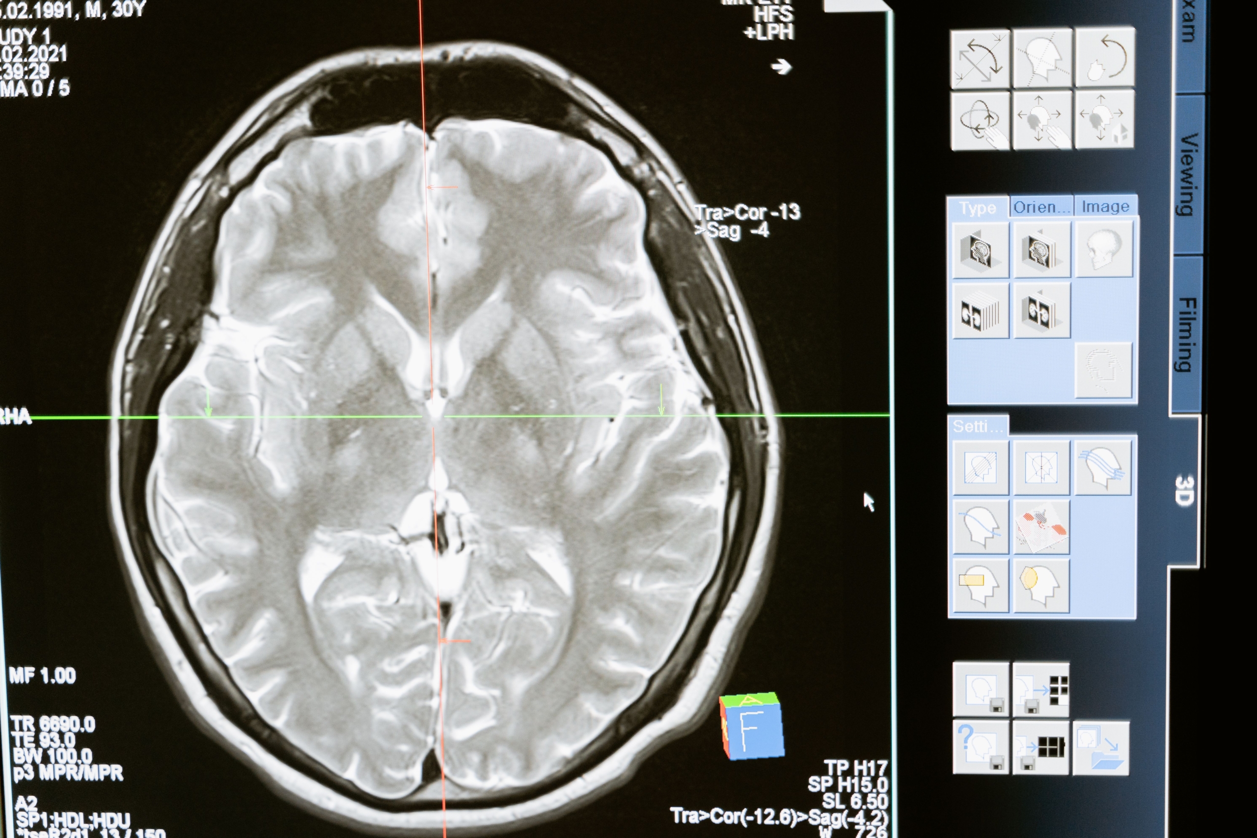
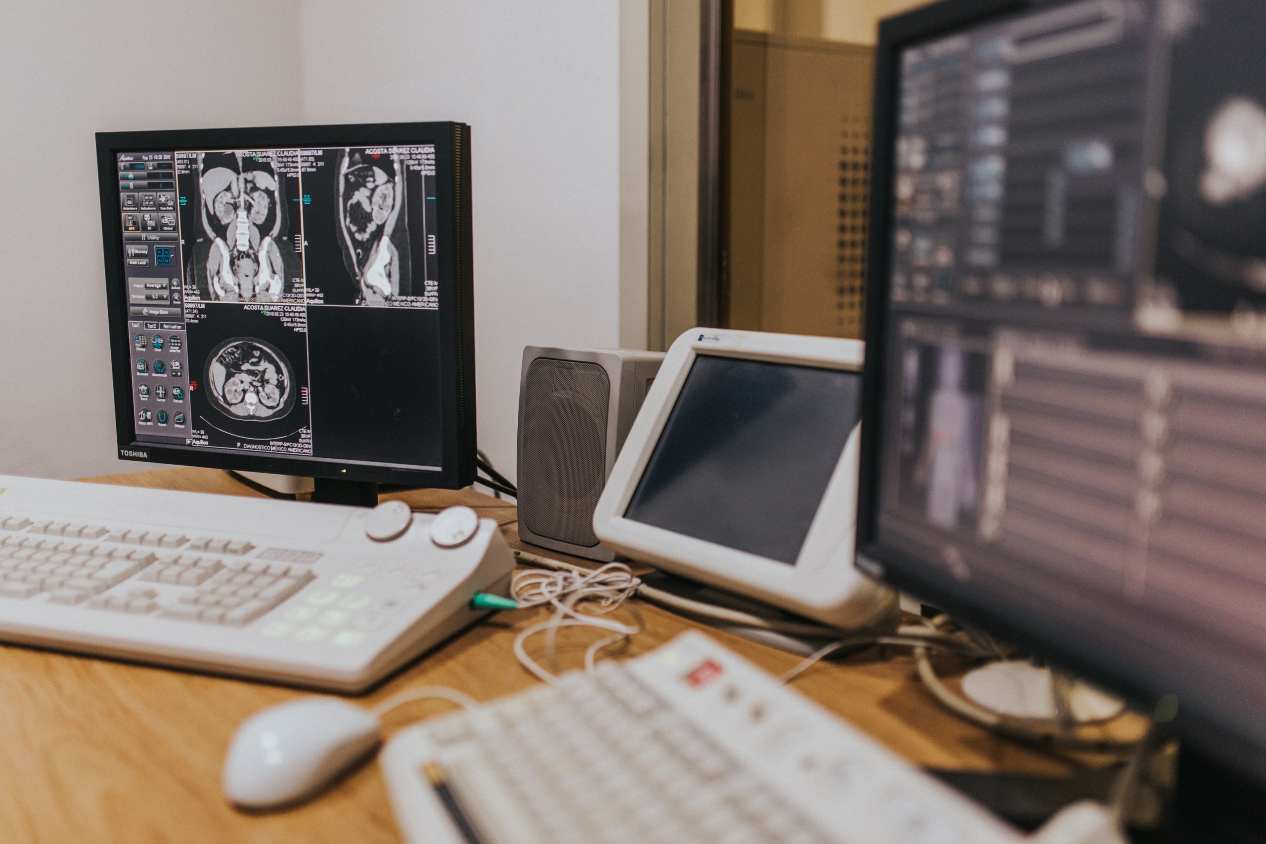
Medical Imaging in Radiology and Artificial Intelligence
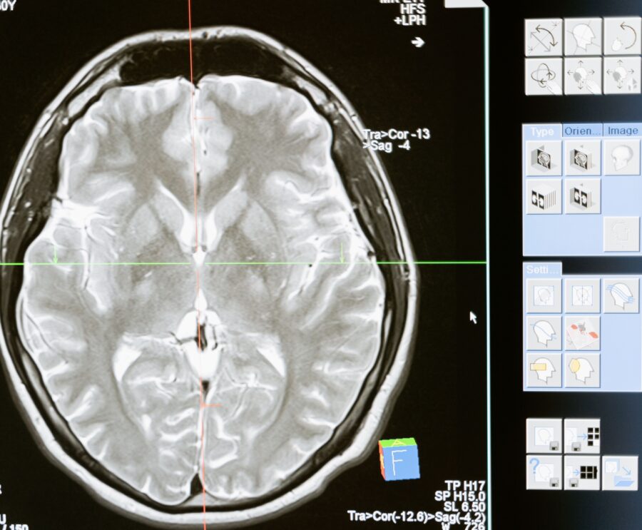
Medical imaging encompasses a range of techniques used to produce visual representations of the inside of the human body to detect, characterize, and monitor diseases. It includes modalities such as conventional X-rays, ultrasound, computed tomography (CT), magnetic resonance imaging (MRI), mammography, and nuclear medicine (PET). Image interpretation has traditionally relied on the radiologist’s expertise, a complex process that can be affected by fatigue, workload, and variability between professionals.
Artificial intelligence adds the ability to automatically analyze large volumes of images and recognize patterns that may sometimes go unnoticed by the human eye. Within AI, it is useful to distinguish between several concepts:
- Artificial intelligence: Systems capable of replicating human abilities such as learning, reasoning, and perception.
- Machine learning: Techniques that identify patterns in data to make decisions without being explicitly programmed to do so.
- Deep learning: Models based on multilayer neural networks that automatically learn relevant features from images. This technology has driven the major advances in AI in radiology.
Radiology is by far the medical specialty with the largest number of approved AI solutions. According to data published through the first quarter of 2026, the US Food and Drug Administration (FDA) had authorized more than 1,500 AI-enabled medical devices, approximately 76% of which were in the field of radiology. Major medical imaging equipment manufacturers such as GE HealthCare, Siemens Healthineers, Philips, and Canon, together with specialized software companies such as Aidoc and Rapid.ai, are leading this market.
Spanish hospitals are no exception to this trend. Quirónsalud already uses algorithms in routine imaging services, from reviewing emergency department X-rays to mammography and cardiac MRI, while Hospital Clínic de Barcelona has begun gradually deploying these tools. In both cases, the underlying idea is the same: technology takes over some of the repetitive work so that radiologists can devote their time to the cases that genuinely require their expertise.
One factor explains the urgency of this transition: the number of images generated per examination has multiplied. Modern CT scanners produce hundreds or even thousands of slices per scan, an amount that is impossible to review with the same level of attention throughout an entire working day. AI is no longer a luxury; it is becoming a response to a problem of scale.
How AI-Powered Medical Imaging Works in Radiology
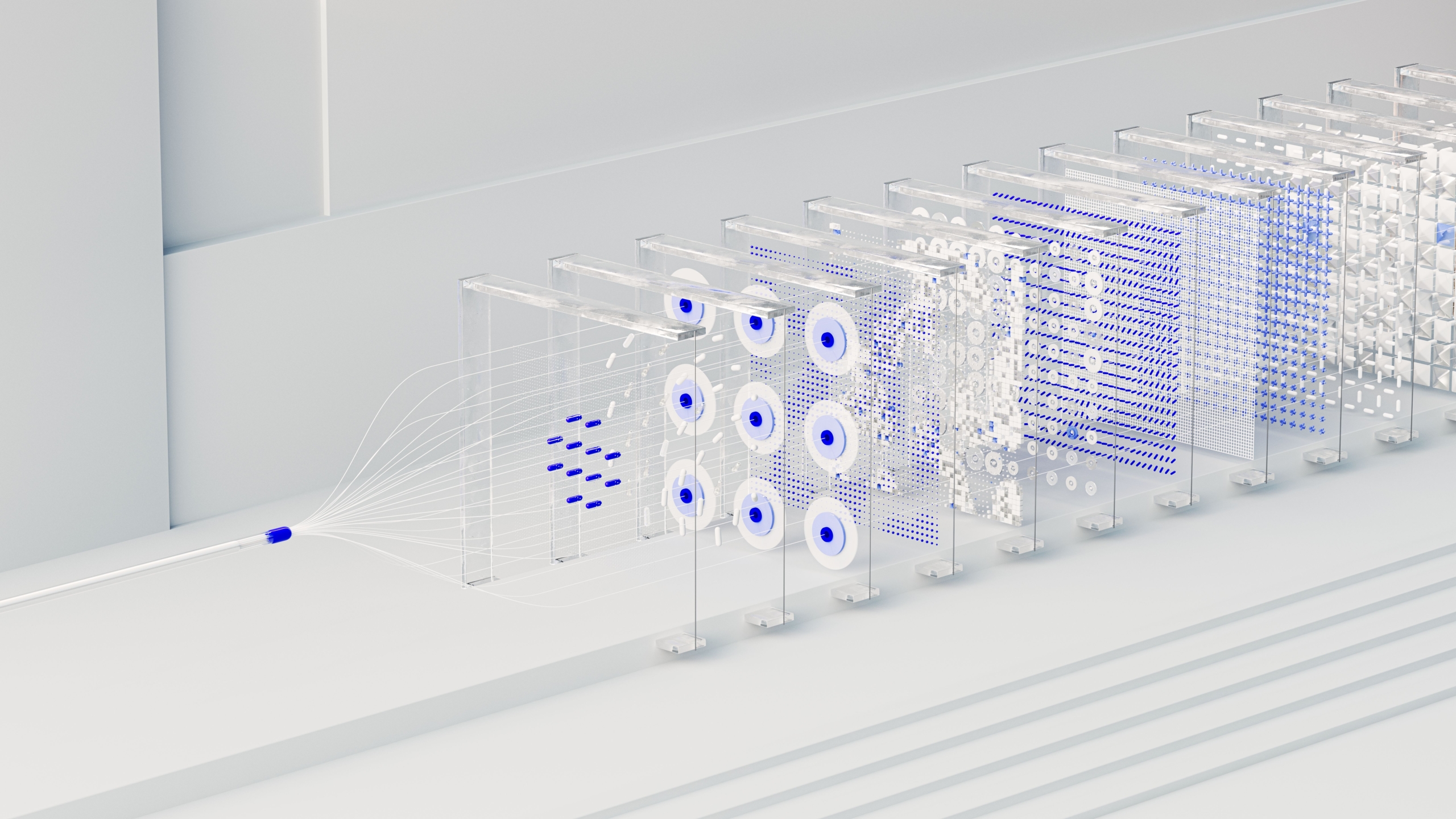
Most AI systems applied to medical imaging are based on convolutional neural networks (CNNs), a type of deep learning model specifically designed to work with images.
Convolutional Neural Networks
One simple way to understand a convolutional neural network is to compare it with how a radiologist learns to interpret an image: first distinguishing contours and densities, then recognizing anatomical structures, and finally identifying what is normal and what is not. The network replicates this process in stages:
- Feature extraction: Small filters move across the entire image and respond to local patterns, such as an edge, a change in intensity, or a grainy texture.
- Downsampling and summarization: The information is condensed to retain the most relevant features, so that the result does not change simply because a lesion is a few millimeters farther to the left or right.
- Decision-making: The final layers combine everything the network has learned and translate it into a specific output, such as a probability of malignancy or the location of a finding.
The more layers the information passes through, the more complex the patterns the network can recognize. It progresses from simple lines to shapes and then to increasingly complex structures.
Model Training
No algorithm inherently knows medicine; it learns from examples. During training, it is presented with large collections of medical studies whose diagnoses have already been confirmed by specialists. Each time the model makes a mistake, it slightly adjusts its internal parameters. After many repetitions, it learns to produce consistently accurate results.
In medicine, obtaining enough well-annotated cases is expensive and time-consuming. For this reason, it is common to reuse models that have already learned to interpret general-purpose images and then specialize them using radiological studies. This strategy is known as transfer learning.
On this basis, AI-powered medical imaging relies on three main tasks:
Classification
The system answers a specific question about an image: Is there a fracture or not? Does the lesion appear benign or suspicious? For well-defined tasks, such as detecting wrist fractures, the literature reports area under the curve (AUC) values of around 0.95.
Detection
Here, it is not enough to determine whether something abnormal is present; the system must also indicate where it is located. The result is usually displayed as a marker or bounding box over the suspicious area. This is particularly useful for locating pulmonary nodules on a CT scan containing hundreds of slices or identifying small brain hemorrhages that might otherwise go unnoticed.
Segmentation
This is the most detailed task: automatically tracing the exact boundaries of an organ or tumor. Performing this manually can take a specialist a considerable amount of time, whereas a model can complete it in seconds. This makes it possible to calculate volumes, compare studies over time, and plan radiotherapy treatments. Architectures such as U-Net have become a standard for this type of task.
In every case, the tool makes a recommendation, and the radiologist makes the decision. AI provides a rapid, tireless second opinion, but the final interpretation, taking each patient’s individual circumstances into account, remains the specialist’s responsibility.
Clinical Applications of medical imaging
Beyond the theory, AI-powered medical imaging is already demonstrating its value in clinical practice. Below are three of the applications with the greatest impact.
Breast Cancer Screening
Mammography screening programs process an enormous number of examinations every year, most of which are normal. In many countries, each mammogram is reviewed by two radiologists. This makes breast cancer screening an ideal setting for AI.
One of the most significant studies to date is the MASAI trial, conducted in Sweden with more than 100,000 women and published in The Lancet. In this trial, the algorithm assigned a risk score to each examination to determine how many radiologists needed to review it. It also highlighted areas requiring closer attention.
After two years of follow-up, compared with conventional screening, the group supported by AI achieved the following results:
- Fewer cancers diagnosed between screening rounds, known as interval cancers (approximately 12% fewer).
- An even greater reduction, of around 27%, in cancer subtypes associated with a poorer prognosis.
- More cancers detected during screening itself, mostly small tumors at an early stage.
- Almost half as many radiologist readings, without an increase in false alarms.
Stroke Detection and Triage
In an ischemic stroke, every minute without treatment results in the loss of millions of neurons. When the cause is a blood clot blocking a major cerebral artery, the most effective treatment is to remove it through a catheter-based procedure called a thrombectomy. How quickly the medical team is mobilized is critical.
In this setting, AI acts as an early warning system: it analyzes CT angiography images as soon as they are acquired and immediately alerts the stroke team if it suspects an arterial occlusion.
The impact of this approach was measured in a randomized trial published in JAMA Neurology, involving 243 patients treated at four hospitals in the United States. With the automated system, the time until thrombectomy began was reduced by an average of approximately 10–11 minutes. This may seem like a small difference, but in the context of stroke, it is clinically significant.
Automated Report Generation
The radiology report is the final product of a radiologist’s work and also one of the most time-consuming tasks. The emergence of language models capable of interpreting images has opened the door to AI generating an initial draft based on the current examination, previous scans, and available clinical information. The specialist then only needs to review, correct, and sign the report.
Models specifically designed for radiology already exist, such as Harrison.Rad 1.5, introduced in 2026 and designed for plain X-rays and mammography. This is still an emerging field, and its results need to be confirmed through independent studies. However, its potential to free up time currently spent on administrative tasks is clear.
Benefits of AI-Powered Medical Imaging
The benefits of AI-powered medical imaging can be seen at three levels: patients, who receive diagnoses sooner and with greater confidence; healthcare professionals, who gain time and support; and the healthcare system, which can handle greater demand with the same resources. Some of the key benefits include:
- Greater diagnostic accuracy: A well-trained algorithm does not become distracted or tired and can identify findings just a few millimeters in size or with very low contrast. It acts as a safety net, reducing the likelihood that something important will go unnoticed.
- Faster diagnoses: By analyzing each examination as soon as it is acquired, AI makes it possible to reorder the workload and prioritize critical cases, such as a brain hemorrhage or pneumothorax, rather than reviewing them in the order they arrive.
- Reduced workload: A significant proportion of medical imaging studies show no abnormalities. If AI can reliably help identify these cases, radiologists can devote more attention to difficult examinations, improving both the quality of their work and their well-being.
- Less variability between professionals: Two specialists may interpret the same image differently, and the same professional may reach different conclusions at the beginning and end of a shift. AI applies the same criteria consistently, helping improve uniformity.
- Objective quantification: Statements such as “the lesion appears slightly larger” can be replaced with precise measurements: millimeters, cubic centimeters, or percentage changes. This makes it easier to determine whether a treatment is working.
- Towards personalized and predictive medicine: Medical images contain information that the human eye cannot quantify. When combined with medical history, laboratory results, or genetic profiles, this information can help predict how a disease may progress or which treatment is most likely to work for an individual patient.
Challenges of AI-Powered Medical Imaging
An algorithm performing well in a scientific paper does not mean it will work equally well in every hospital. There is a long journey between research and everyday clinical use. The main challenges include:
- Data quality and representativeness: A model is only as good as the examples it learned from. If its training data predominantly include patients with a particular profile, images from a single manufacturer, or cases from one hospital, its performance may deteriorate when used in a different environment.
- Validation in real-world settings: Before relying on a tool, it is advisable to test it using the hospital’s own imaging studies and continue monitoring its performance over time, as equipment, protocols, and patient populations change.
- Explainability: A simple “yes” or “no” is not enough for a physician; they need to understand the basis for the machine’s conclusion. This is why tools that highlight the image regions influencing a result and indicate the level of confidence are becoming increasingly important.
- Integration into clinical workflows: If radiologists have to open another application, wait for results, or manually copy data, the tool will eventually go unused. AI should be integrated into existing image viewers and systems used in daily practice and should be compatible with equipment from different manufacturers.
- Ethical and privacy considerations: Medical images contain highly protected health data. Another unresolved question also arises: who is responsible if a system error contributes to an incorrect diagnosis?
- Professional training: Using AI effectively requires an understanding of its limitations. Radiologists do not need to be programmers, but they must know how to interpret algorithm performance metrics, recognize common failure modes, and avoid relying on AI blindly.
Current Regulations and Standards: The Situation in Spain
From a legal perspective, software that helps diagnose diseases using medical images is not just any software: it is a medical device. For this reason, in the European Union, it must comply with two major regulatory frameworks that complement each other.
Medical Device Regulation (MDR)
Regulation (EU) 2017/745 establishes the safety and performance requirements that medical devices must meet. For diagnostic software, this involves obtaining CE marking following assessment by an independent notified body.
European Artificial Intelligence Act (AI Act)
Regulation (EU) 2024/1689 is the world’s first comprehensive law on artificial intelligence. It entered into force in August 2024, with requirements being implemented in phases. It classifies as “high-risk” AI systems integrated into medical devices that require third-party conformity assessment, which includes most radiology AI tools. The implementation timeline for these systems extends through 2027.
In practice, among other requirements, the regulation obliges manufacturers of these systems to implement:
- A risk management system that remains active throughout the product’s time on the market (Article 9).
- Relevant and representative datasets that are reviewed for potential bias (Article 10).
- A design that enables human oversight, allowing healthcare professionals to interpret the results and decide not to follow the system’s recommendations (Article 14).
- Clear instructions for use, automatic event logging, and post-market performance monitoring (Articles 12, 13, and 72).
Healthcare facilities using these tools also have responsibilities: they must use them according to the manufacturer’s instructions, assign their supervision to qualified personnel, and report any problems they identify.
The Situation in Spain
Two main organizations are involved in Spain: the Spanish Agency of Medicines and Medical Devices (AEMPS), which is responsible for medical devices, and the Spanish Agency for the Supervision of Artificial Intelligence (AESIA), based in A Coruña, which is responsible for overseeing the implementation of the European AI regulation. This framework is complemented by the General Data Protection Regulation (GDPR) and Spain’s Organic Law on Personal Data Protection and Guarantee of Digital Rights (LOPDGDD), which govern the processing of health information.
In practice, radiology is the hospital department making the greatest progress in AI adoption in Spain. Hospitals such as La Paz in Madrid, Hospital Clínic in Barcelona, and Quirónsalud facilities already have projects underway. Nevertheless, the landscape remains uneven: adoption largely depends on the initiatives and resources available at each hospital or within each autonomous community, and there is still room for a more coordinated nationwide strategy.

Few areas of medicine demonstrate the value of artificial intelligence as clearly as medical imaging. Applications ranging from mammography screening to early stroke alerts and automated report writing show that AI is no longer just a promise; it is a tool delivering measurable results.
The challenge now is to bring it from the laboratory into clinical practice safely and reliably: validating it with local data, integrating it into everyday workflows, complying with the new regulations, and training healthcare professionals. Throughout this process, one principle must remain clear: the radiologist has the final say.
As these technologies continue to evolve, AI-powered medical imaging will become one of the pillars of a more efficient, personalized, and patient-centered approach to medicine.
Would you like to learn more about computer vision and applied AI? Get in touch with us!